
Telogen effluvium diagnosis
Reviewed by
Steven P., FAAD
Board-certified dermatologist
Updated on
Reviewed for accuracy
Table of Contents
Dermatologists diagnose telogen effluvium by matching your sudden diffuse shedding and hair‑pull test (excess telogen club hairs) with a detailed history and targeted labs. They’ll time shedding to triggers 2-3 months earlier (illness, surgery, childbirth, crash diet, major stress), review drugs, menses, and diet, examine scalp density and shaft caliber, and order ferritin, iron studies, thyroid tests, vitamin D, B12, and zinc. You’ll also see how this diagnosis shapes prognosis and treatment decisions.
Telogen Effluvium Diagnosis Summary
- Diagnosis is clinical: sudden diffuse shedding 2-3 months after a trigger (illness, stress, childbirth, surgery, diet change) with overall reduced volume, not bald patches.
- Scalp exam shows non‑scarring loss with preserved follicles, uniform shaft caliber, and no shiny atrophic areas, distinguishing from scarring and androgenetic alopecia.
- Hair‑pull test and standardized wash test quantify active shedding; >10-15% telogen club hairs or >100-150 hairs/day support telogen effluvium.
- Targeted labs (ferritin/iron studies, TSH/free T4, vitamin D, B12, zinc, CBC, CMP) identify common reversible triggers like iron deficiency or thyroid disease.
- Trichoscopy and, when uncertain, scalp biopsy confirm increased telogen follicles and exclude alopecia areata, scarring alopecias, or miniaturizing androgenetic alopecia.
How is Telogen Effluvium Diagnosed?
When specialists diagnose telogen effluvium, they systematically combine a detailed history, targeted scalp and hair examination, and, when indicated, focused laboratory testing rather than relying on a single test. Your clinician asks when shedding began, whether it followed illness, surgery, childbirth, rapid weight change, medication adjustments, or intense workplace stress. They’ll explore psychological impact, sleep, diet, and any seasonal patterns in shedding.
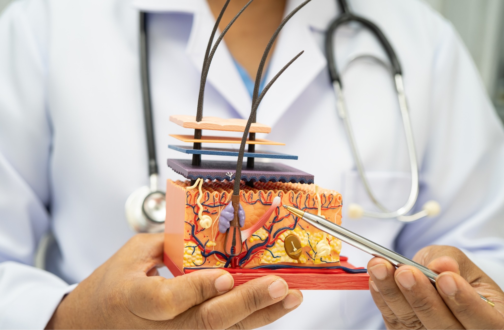
Next, they examine your scalp density, part width, and hair shaft caliber, evaluating for miniaturization that would suggest androgenetic alopecia instead. They evaluate scalp hygiene, looking for scale, erythema, or folliculitis that might contribute to shedding, and review your haircare routines, including traction hairstyles, chemical treatments, and heat exposure.
A gentle hair-pull test and review of shed hairs (telogen club hairs vs broken shafts) help confirm diffuse, non-scarring loss. This structured process validates your experience and anchors it in clear, observable findings.
Blood Tests for Telogen Effluvium
When your clinician suspects telogen effluvium, they typically order targeted blood tests to identify systemic contributors that disrupt the hair cycle.
You’ll usually be evaluated for low ferritin/iron stores, thyroid dysfunction, and micronutrient deficiencies such as vitamin D, vitamin B12, and zinc, all of which have documented associations with increased telogen shedding.
Low Ferritin or Iron and Telogen Effluvium
Recognizing the link between iron status and telogen effluvium is essential because low ferritin—your body’s main iron storage protein—is one of the most common and treatable contributors to diffuse hair shedding. Your clinician should check serum ferritin, iron, total iron binding capacity, and transferrin saturation to assess storage and transport.
In practice, many hair specialists use higher ferritin thresholds (often ≥40-70 ng/mL) for optimal hair cycling, even when labs label lower values “normal.” Heavy menstrual bleeding, frequent blood donation, restrictive diets, and gastrointestinal losses commonly push ferritin below these ranges. You may also experience low ferritin despite normal hemoglobin.
When deficiency’s confirmed, targeted iron supplements, timed around hepcidin regulation and food interactions, can safely restore iron stores and support regrowth.
Thyroid Issues and Telogen Effluvium
Although thyroid disease often gets overlooked in diffuse shedding, your clinician should systematically screen for it because both hypothyroidism and hyperthyroidism can trigger telogen effluvium by disrupting the hair cycle’s anagen (growth) phase. You’ll typically have TSH, free T4, and sometimes free T3 checked, with additional tests guided by history and symptoms.
Because thyroid autoimmunity is common in people with hair loss, your clinician may order anti‑TPO and anti‑TG antibodies, especially if you have subclinical hypothyroidism, fluctuating TSH, or a family history of autoimmune disease. In the postpartum period, targeted testing can uncover postpartum thyrotoxicosis as a reversible driver of shedding.
Your provider will also review thyroid medication dosing, prior radioactive iodine or surgery, and iodine intake to optimize thyroid status and support regrowth.
Vitamin D Deficiency
Beyond thyroid function, your clinician will usually assess vitamin D status because low 25‑hydroxyvitamin D [25(OH)D] appears more common in patients with telogen effluvium and other non‑scarring alopecias than in controls.
Your blood test typically targets a level above 30 ng/mL, though laboratories differ. Deficiency doesn’t prove causation, but it’s a modifiable factor you and your team can address together.
Key elements your clinician may review:
-
Seasonal variation: reduced sunlight exposure in winter often lowers 25(OH)D.
-
Dietary sources: limited intake of fatty fish, fortified dairy, or eggs can contribute.
-
Genetic predisposition: variants in vitamin D related genes may reduce bioavailability.
-
Supplement dosing: tailored cholecalciferol regimens, with monitoring, help avoid under‑ or over‑replacement.
Vitamin B12 Deficiency
Because vitamin B12 participates in DNA synthesis and erythropoiesis, clinicians often include a serum B12 level when evaluating diffuse shedding or suspected telogen effluvium, especially if you also report fatigue, paresthesias, glossitis, or anemia. You’ll usually have total serum B12 checked, but your clinician may add methylmalonic acid and homocysteine to uncover “functional” deficiency and clarify methylation status.
They’ll ask about dietary sources (meat, fish, eggs, dairy) and absorption factors such as pernicious anemia, gastric surgery, metformin, or proton‑pump inhibitors.
Untreated deficiency can cause progressive neurological symptoms like numbness, burning feet, gait disturbance, cognitive changes that may accompany hair shedding and make you feel alarmingly “not yourself.”
Evidence‑based supplementation strategies include high‑dose oral cyanocobalamin or methylcobalamin, or intramuscular injections when absorption is impaired.
Zinc Deficiency
Zinc status is another laboratory checkpoint when you’re being evaluated for telogen effluvium, since zinc is essential for keratinocyte proliferation, protein synthesis, and normal hair‑follicle cycling. Your clinician will usually order serum zinc and related Zinc biomarkers, sometimes alongside serum copper to assess the Zinc-to-copper ratio, which helps detect marginal deficiency.
-
Testing focus: Low serum zinc, with or without low alkaline phosphatase, supports zinc deficiency contributing to diffuse shedding.
-
Context matters: Inflammation, hypoalbuminemia, and oral contraceptives can alter Zinc absorption and circulating levels.
-
Treatment: Carefully dosed Zinc supplementation may normalize shedding over several months, especially when integrated with overall nutritional support.
-
Safety: Your team will monitor for Zinc toxicity and secondary copper depletion, adjusting doses to keep you safely within physiological ranges.
Key Symptoms and Patterns Dermatologists Look for
When dermatologists evaluate suspected telogen effluvium, they focus on specific patterns of hair shedding and scalp findings rather than vague “thinning.” They look for a history of increased daily hair loss (often >100 to 150 hairs/day), sudden onset typically 2-3 months after a trigger (such as illness, surgery, or childbirth), and diffuse shedding from the entire scalp rather than well-defined bald patches.
You’ll usually notice extra hair on your pillow, in the shower, and when you gently tug a small bundle. Dermatologists also ask if shedding follows seasonal patterns, flares with emotional triggers, or coincides with starting or stopping hormonal contraception. They pay attention to whether your scalp looks normal in color and texture, which supports a diagnosis of telogen effluvium rather than inflammatory disease.
Emerging research explores how alterations in the scalp microbiome and measurable stress biomarkers might parallel these shedding patterns, reinforcing that your experience is biologically real, not “in your head.”
Medical History and Physical Examination in Diagnosis
Although telogen effluvium is often obvious to patients, dermatologists rely on a structured medical history and targeted physical examination to confirm the diagnosis and rule out mimicking conditions. Your visit typically starts with a detailed timeline of shedding onset, peak, and stabilization, linked to triggers such as illness, surgery, childbirth, crash dieting, or major stressors.
Dermatologists then systematically evaluate:
-
Medication review – You’ll go through prescription, over‑the‑counter, and supplement use, focusing on drugs known to precipitate shedding (retinoids, anticoagulants, β‑blockers, thyroid agents).
-
Hormonal evaluation clues – Menstrual patterns, pregnancies, perimenopause, androgen‑related symptoms guide later lab choices.
-
Autoimmune screening cues – Joint pain, rashes, thyroid symptoms, or family history raise suspicion for systemic disease.
-
Psychosocial assessment – You’re invited to share stressors, mood changes, and body‑image concerns to contextualize triggers and support needs.
Physical examination documents scalp coverage, part width, hair shaft caliber, and pattern symmetry; a scalp biopsy is reserved for diagnostic uncertainty.
Diagnostic Tests: Pull Test, Wash Test, and Trichoscopy
Objective tests like the hair‑pull test, standardized wash test, and trichoscopy translate your shedding history into measurable findings and help distinguish telogen effluvium from other alopecias.
In the hair‑pull test, your clinician gently tugs a bundle of hairs; extracting more than 10-15% telogen‑club hairs suggests active shedding, often after stress testing events, drug triggers, illness, or childbirth.
The standardized wash test quantifies daily loss: you avoid shampooing for five days, then wash over a gauze or filter; counting the shed hairs, and classifying them by roots and shaft diameter, helps separate telogen effluvium from androgenetic alopecia or seasonal shedding patterns.
Trichoscopy (dermoscopy of the scalp) lets your provider view the hair cycle in real time, evaluating shaft diameter diversity, empty follicles, and regrowth stubble.
When patterns remain unclear, a targeted scalp biopsy can confirm increased telogen follicles and exclude scarring or inflammatory alopecias.
Telogen Effluvium ICD-10
Because telogen effluvium is a clinical and exclusion-based diagnosis, your clinician usually orders targeted laboratory tests to uncover correctable triggers and then documents the condition using the appropriate ICD‑10 code. You’ll typically see hair loss recorded under codes such as L65.9 (nonscarring hair loss, unspecified), sometimes alongside codes for iron deficiency, thyroid disease, or recent childbirth to capture postpartum onset.
Common lab panels aim to connect your symptoms with disruptions in the hair cycle, systemic inflammation, or a silent genetic predisposition:
-
Nutritional / endocrine tests – CBC, ferritin, iron studies, vitamin D, B12, TSH, free T4.
-
Metabolic and inflammatory markers – CMP, CRP, sometimes cortisol or other stress biomarkers.
-
Reproductive / postpartum context – hCG, prolactin, or documentation of recent delivery.
-
Adjunctive research-focused tests – exploration of the scalp microbiome or genomic panels in specialized settings.
Telogen Effluvium vs. Other Hair Loss Conditions
TE often follows a systemic trigger classically stress alopecia after major illness, childbirth, crash dieting, or psychosocial stressors, but also medication induced from agents like retinoids, anticoagulants, or certain antidepressants.
In contrast, an androgenic pattern (androgenetic alopecia) shows miniaturization in characteristic frontal and vertex distributions, not uniform shedding.
You also need to distinguish TE from scarring alopecia, where inflammation destroys follicles, leaving shiny, atrophic skin, and from autoimmune alopecia, such as alopecia areata, which produces sharply demarcated patches or ophiasis patterns, often with exclamation‑point hairs at the margins.
What a Diagnosis Means for Prognosis and Treatment Planning
Once you establish that diffuse shedding represents telogen effluvium rather than another alopecia, the diagnosis immediately frames both prognosis and the therapeutic strategy.
You’re no longer asking “what’s happening?” but “how do we guide recovery?” With TE, the long term outlook is usually favorable because follicles remain viable; the focus shifts to identifying triggers, mapping a realistic treatment timeline, and protecting your mental health along the way.
Key elements of a TE‑specific plan include:
-
Prognosis – Estimate regrowth based on trigger removal, chronicity, and comorbid conditions; discuss expected shedding and density milestones.
-
Therapeutic targeting – Correct nutritional, endocrine, or medication-related causes; individualize adjuncts like minoxidil.
-
Psychological impact – Screen for anxiety, body‑image distress, and social withdrawal; normalize your experience and offer support options.
-
Relapse prevention and cost planning – Build monitoring intervals, labs, and lifestyle changes into follow‑up, while structuring affordable, stepwise interventions.
TE Diagnosis FAQs
Can Telogen Effluvium Be Diagnosed Through Online Photos or Telemedicine Consultations?
You can’t reliably confirm it through photos alone; telemedicine limitations, variable image quality, and patient privacy concerns require in‑person or remote trichoscopy, standardized scalp photographs, labs, and clear followup logistics with your clinician for a confident diagnosis.
Does Stress-Related Telogen Effluvium Require Different Diagnostic Tests Than Other Triggers?
You usually won’t need different core tests, but stress‑related telogen effluvium may justify stress biomarkers, psychosocial assessment, autonomic testing, and hormonal modulation review, guiding tailored behavioral interventions while still using standard labs and scalp examination.
How Long After Shedding Starts Should I Wait Before Seeking Diagnostic Evaluation?
You shouldn’t let your hair down on this one—don’t use a long wait period. When early signs persist beyond 4-6 weeks of shedding, that’s ideal consult timing; this diagnostic window helps determine when to seek targeted evaluation.
Can Nutritional Supplements Affect the Accuracy of Blood Tests for Telogen Effluvium?
Yes, supplements can skew results. Biotin masking alters thyroid and hormone assays; vitamin interference and herbal supplements affect liver, kidney, and coagulation panels; iron supplementation distorts ferritin/iron indices; excess fat soluble vitamins (A, D, E, K) misleads nutritional assessment.
Is a Scalp Biopsy Ever Necessary for Diagnosing Chronic Telogen Effluvium?
You almost never need a scalp biopsy, but when clinical doubt screams, you’ll use it for invasive confirmation, evaluating diagnostic utility, histopathology features, and multiple sampling sites, so you’re absolutely sure it’s chronic telogen effluvium and not alone guessing.
Reviewed by
Steven P., FAAD
Board-certified dermatologist
Updated on
Reviewed for accuracy