
Telogen Effluvium Pictures
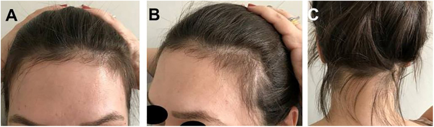
Telogen effluvium pictures provide a visual reference for what this type of hair loss looks like in real life. These images can help people compare their own hair changes to typical patterns seen in others with the condition. Common features include overall thinning, a wider part, and more visible scalp, especially under bright lighting or when the hair is wet.
While photos can be useful for tracking changes over time, it is important to remember that every individual’s hair density and pattern are unique. Lighting, hair color, and styling can also affect how thinning appears in pictures. Photos should be used as a guide, not a substitute for professional evaluation.
Diffuse Thinning
Part Widening
No Bald Spots
Regrowth Visible
Why Photos are Helpful
Photos allow individuals to objectively monitor changes in their hair over time. By taking regular pictures of the scalp, especially the part, crown, and temples, it becomes easier to notice subtle changes that may not be obvious day to day. Comparing before and after photos can also help distinguish between ongoing shedding and new hair growth.
For healthcare providers, photos can supplement the clinical history and physical exam. They provide a visual timeline of hair loss and regrowth, which can be valuable for diagnosis and treatment planning. However, a diagnosis of telogen effluvium should not be made based on photos alone. Always consult a healthcare professional for an accurate assessment.
Common Visual Signs
Telogen effluvium most often presents with diffuse thinning, meaning hair loss is spread evenly across the scalp. The hair part may appear wider, and more scalp may be visible, especially under certain lighting. These changes can be subtle at first but become more noticeable as shedding continues.
Unlike some other forms of hair loss, telogen effluvium does not typically cause bald patches or scarring. The overall hair density decreases, but the hairline usually remains intact. Key areas to examine in photos include the crown, temples, and along the part.
Diffuse Thinning
Diffuse thinning refers to a general reduction in hair density across the entire scalp. In telogen effluvium, this thinning is usually even, without distinct patches. Photos often show less volume and a more see-through appearance, especially when hair is pulled back or parted.
This type of thinning can make the scalp more visible, particularly in bright light or when the hair is wet. Diffuse thinning is a hallmark of telogen effluvium and helps distinguish it from conditions like alopecia areata, which causes patchy hair loss.
Part Widening
One of the earliest and most noticeable signs in telogen effluvium pictures is widening of the hair part. As hair density decreases, the part line appears broader and more scalp shows through. This is especially apparent in overhead or top-down photos.
Part widening is often most visible in women, but it can occur in anyone with telogen effluvium. Comparing photos taken weeks or months apart can help track changes in the width of the part and overall scalp coverage.
Changes at the Crown and Temples
The crown and temples are areas where thinning may be particularly noticeable in telogen effluvium. Photos may show increased scalp visibility at the crown, especially when hair is parted or styled away from the area. The temples may also appear thinner, with less fullness at the sides of the forehead.
These changes are typically subtle and diffuse, not sharply defined. The hairline usually remains unchanged, which helps differentiate telogen effluvium from other types of hair loss that affect the frontal hairline or cause recession.
Before and After Telogen Effluvium Photos
Before and after photos are a valuable tool for tracking the progression and recovery of telogen effluvium. By comparing images taken before hair shedding began with those taken during and after the episode, individuals can see changes in hair density, part width, and scalp visibility.
In before photos, the hair often appears fuller, with a narrow part and less visible scalp. After telogen effluvium sets in, photos may show increased thinning, a wider part, and more scalp showing through. As regrowth occurs, new short hairs may be visible, and overall density gradually improves.
How to Recognize Regrowth
Recognizing regrowth in telogen effluvium pictures involves looking for specific signs. New hair growth often appears as short, fine, and sometimes wispy hairs along the hairline, temples, and part. These new hairs may stand out from the rest of the hair due to their length and texture.
Over time, regrowth becomes more noticeable as these short hairs grow longer and blend in with the surrounding hair. Photos taken at regular intervals can help document this process. It is important to remember that regrowth may take several months, and the appearance of new hairs is a positive sign of recovery.
What Does Early Telogen Effluvium Look Like?
In the early stages, telogen effluvium may be difficult to spot in photos. The first sign is often an increase in hair shedding, which may not immediately result in visible thinning. As the condition progresses, subtle changes such as a slightly wider part or less volume may become apparent.
Early telogen effluvium pictures may show minimal changes, but careful comparison over time can reveal gradual thinning. Taking consistent photos in similar lighting and angles can help detect these early changes. For those concerned about new hair loss, early documentation can be valuable for both self-monitoring and discussions with healthcare providers.
Frequently Asked Questions
Can telogen effluvium cause bald spots?
Telogen effluvium typically causes diffuse thinning rather than bald spots. The hair loss is spread evenly across the scalp, and patchy bald areas are not a common feature. If bald spots are present, another condition such as alopecia areata may be involved.
How can I tell if my hair loss is telogen effluvium?
Telogen effluvium usually presents as increased shedding and overall thinning, with a wider part and more visible scalp. Photos can help track these changes, but diagnosis should be confirmed by a healthcare provider.
How long does it take to see regrowth?
Regrowth after telogen effluvium can begin once the underlying trigger is resolved. Short, fine hairs may become visible within a few months, and full recovery may take several additional months. The timeline varies for each person.
Will my hair look normal again?
Most people with telogen effluvium experience full regrowth once the cause is addressed. Hair density gradually returns to normal, and the part narrows as new hair grows in. Recovery times can vary, but permanent damage is rare.
Is it normal to see more scalp?
Yes, increased scalp visibility is a hallmark of telogen effluvium. Thinning is usually even, making the scalp more noticeable, especially along the part and crown. This is a common feature seen in telogen effluvium photos.
Are before and after photos reliable?
Before and after photos can be helpful for tracking changes in hair density and regrowth, but they should be taken in consistent lighting and angles for accuracy. Photos are a useful tool but should not replace professional evaluation.
Can photos help a doctor diagnose me?
Photos can support a doctor’s assessment by providing a visual record of hair changes over time. However, diagnosis of telogen effluvium requires a full clinical evaluation and consideration of medical history. Photos alone are not sufficient for diagnosis.
Fact Checked
Updated: December 30, 2025
Reviewed for accuracy against authoritative clinical sources and peer reviewed dermatology references. Educational content only.
Quality Controlled
We use a structured editorial process focused on clarity, accuracy, and alignment with current clinical understanding. This content is not a substitute for professional medical advice.
Editorial Policy
Clinical Sources